Tendon Diagram - Achilles Tendon Rupture And Achilles Tendonitis Treatment / The achilles tendon is the largest.
byAdmin-
0
Tendon Diagram - Achilles Tendon Rupture And Achilles Tendonitis Treatment / The achilles tendon is the largest.. Patellar tendonitis is a knee injury affecting the patella tendon. When the biceps contracts, it pulls the forearm up and rotates it outward. It attaches to the wrist bone, the pisiform, and as well as the 5th hand bone. Bones, muscles, tendons and nerves which will each give slightly different foot pain symptoms. A tendon, also known as a sinew, is a fibrous tissue that helps to facilitate this movement.
Your biceps tendons attach the biceps muscle to bones in the shoulder and in the elbow. Intermediate back muscles and c. This sudden, tight, intense lower leg pain is sometimes called a charley horse. Calcific tendonitis (or tendinitis) occurs when calcium deposits bu. There are a whole range of structures e.g.
Knee Anatomy from embed.widencdn.net This sudden, tight, intense lower leg pain is sometimes called a charley horse. The wrist is actually a collection of many bones and joints. Jul 05, 2018 · the foot diagram has a complex structure made up of bones, ligaments, muscles, and tendons. The achilles tendon is the strongest and largest tendon in the body. Attaches the calf muscles to the calcaneus, most important muscles for running, jumping, walking etc. The tendon travels along the inside of the forearm on the side of the small finger and crosses the wrist. The achilles tendon is the largest. Intermediate back muscles and c.
A typical tendon organ in limb muscles has an ending of about 0.5.
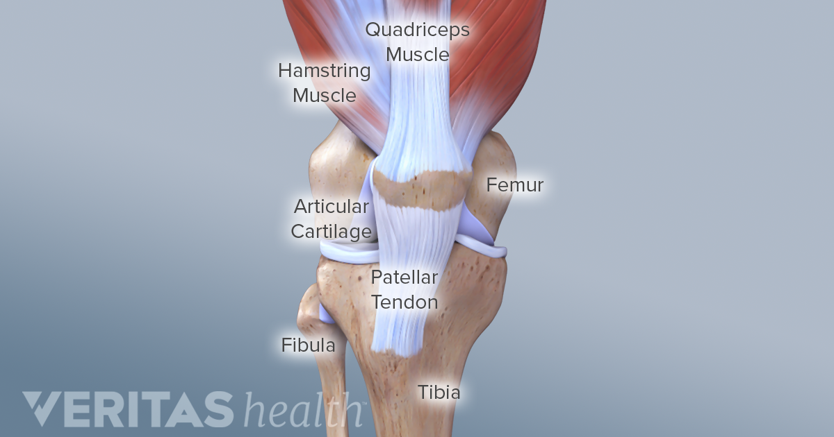
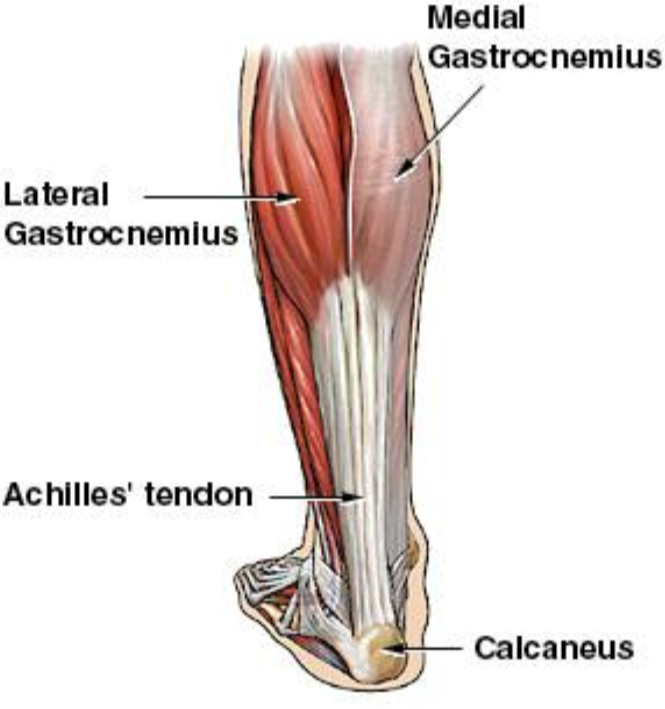
A tendon is a band of tissue that connects a muscle to a bone. Intermediate back muscles and c. This important tendon in the back of the calf and ankle connects the plantaris, gastrocnemius, and soleus muscles to. Allows the action of raising the foot. A typical tendon organ in limb muscles has an ending of about 0.5. You can see a diagram of the achilles tendon below. Tendons are found throughout the body, from the head and neck all the way down to the feet. It attaches to the wrist bone, the pisiform, and as well as the 5th hand bone. In the back and elsewhere in the body, tendons attach muscles to bones. Its muscle belly is in the forearm. The extensor tendon compartments of the wrist are six tunnels which transmit the long extensor tendons from the forearm into the hand. There are a whole range of structures e.g. Ligaments join the knee bones and provide stability to the knee:
The achilles tendon is also called the calcaneal tendon. Tendons, located at each end of a muscle, attach muscle to bone. Attaches the calf muscles to the calcaneus, most important muscles for running, jumping, walking etc. The tendon is firmly connected to muscle fibres at one end and to components of the bone at its other end. A tendon, also known as a sinew, is a fibrous tissue that helps to facilitate this movement.
14 205 Tendon Stock Photos Pictures Royalty Free Images Istock from media.istockphoto.com Hochwertige kletterseile für dein outdoor abenteuer! A typical tendon organ in limb muscles has an ending of about 0.5. The tendon runs down the back of your lower leg from the back of the knee to the heel. Foot anatomy diagram, foot joint diagram, foot sprain diagram, foot tendons and ligaments pain, leg tendon diagram, peroneal tendonitis, foot, foot anatomy diagram, foot joint diagram, foot sprain diagram, foot tendons and ligaments pain, leg tendon diagram, peroneal tendonitis. Tendons attach muscles to bones. Lower back muscle diagram anatomy does degenerative disc disease affect the lower back muscle? Attaches the calf muscles to the calcaneus, most important muscles for running, jumping, walking etc. The anterior cruciate ligament prevents the femur from sliding backward on the tibia (or the tibia sliding forward on the femur).
One peroneal tendon attaches to the outer part of the midfoot, while the other tendon runs under the foot and attaches near the inside of the arch.
9 photos of the foot tendons and ligaments diagram. Tendons are found throughout the body, from the head and neck all the way down to the feet. These structures work together to support the body, enable a range of movements, and send messages from the brain to. A muscle's origin is where a tendon attaches it to the *less* movable bone. This sudden, tight, intense lower leg pain is sometimes called a charley horse. Ligaments join the knee bones and provide stability to the knee: The extensor tendon compartments of the wrist are six tunnels which transmit the long extensor tendons from the forearm into the hand. The two peroneal tendons in the foot run side by side behind the outer ankle bone. Human hand tendon diagram (page 1) hand tendons diagram muscle blank drawing these pictures of this page are about:human hand tendon diagram the golgi tendon organ. Tendon, tissue that attaches a muscle to other body parts, usually bones.tendons are the connective tissues that transmit the mechanical force of muscle contraction to the bones; There are a whole range of structures e.g. The achilles tendon is the strongest and largest tendon in the body. Tendons, located at each end of a muscle, attach muscle to bone.
The achilles tendon is a tough band of fibrous tissue that connects the calf muscles to the heel bone (calcaneus). 9 photos of the foot tendons and ligaments diagram. The achilles tendon is the largest. Attaches the calf muscles to the calcaneus, most important muscles for running, jumping, walking etc. Jul 05, 2018 · the foot diagram has a complex structure made up of bones, ligaments, muscles, and tendons.
Achilles Tendon Rupture Core Em from coreem.net A tendon is a band of tissue that connects a muscle to a bone. Ab 50€ portofrei, versand innerhalb 24h, 100 tage retoure, über 1 mio. The achilles tendon is the strongest and largest tendon in the body. This diagram depicts human anatomy tendons and ligaments.human anatomy diagrams show internal organs, cells, systems, conditions, symptoms and sickness information and/or tips for healthy living. Jul 05, 2018 · the foot diagram has a complex structure made up of bones, ligaments, muscles, and tendons. In the back and elsewhere in the body, tendons attach muscles to bones. Foot anatomy diagram, foot joint diagram, foot sprain diagram, foot tendons and ligaments pain, leg tendon diagram, peroneal tendonitis, foot, foot anatomy diagram, foot joint diagram, foot sprain diagram, foot tendons and ligaments pain, leg tendon diagram, peroneal tendonitis. These structures work together to support the body, enable a range of movements, and send messages from the brain to.
Possibly the most important tendon in terms of mobility is the achilles tendon.
Ab 50€ portofrei, versand innerhalb 24h, 100 tage retoure, über 1 mio. Allows the action of raising the foot. Tendons are found throughout the body, from the head and neck all the way down to the feet. The tendon travels along the inside of the forearm on the side of the small finger and crosses the wrist. This important tendon in the back of the calf and ankle connects the plantaris, gastrocnemius, and soleus muscles to. A typical tendon organ in limb muscles has an ending of about 0.5. It attaches to the wrist bone, the pisiform, and as well as the 5th hand bone. Included within the chart are gorgeous illustrations of the pelvic diaphragm, sphincter muscles, gluteus maximus muscles, and over a dozen more. This diagram depicts human anatomy tendons and ligaments.human anatomy diagrams show internal organs, cells, systems, conditions, symptoms and sickness information and/or tips for healthy living. Hochwertige kletterseile für dein outdoor abenteuer! A typical tendon organ in limb muscles has an ending of about 0.5 mm in length. One peroneal tendon attaches to the outer part of the midfoot, while the other tendon runs under the foot and attaches near the inside of the arch. 17 photos of the diagram of shoulder muscles and tendons.